In April 2022, ConceptD 7 SpatialLabs™ Edition “CN715-73G-SL76Z” laptop was released for sale to the public. Kanagawa Dental University, an educator in cutting-edge dentistry, and Kanagawa Dental University Hospital, which promotes digital transformation (DX) in dentistry, were early in implementing the state-of-the-art machine. Such DX activities aid the development of three-dimensional applications, which are being advanced through collaboration among professionals. The involvement of dentists, dental surgeons, dental technicians, and VR/AR specialists is essential for products such as the ConceptD 7 SpatialLabs™ Edition to push the boundaries of dentistry.
Interview with Professor Tomoaki Itamiya: He is responsible for the implementation of the ConceptD 7 SpatialLabs™
The application and development of naked-eye stereoscopic vision
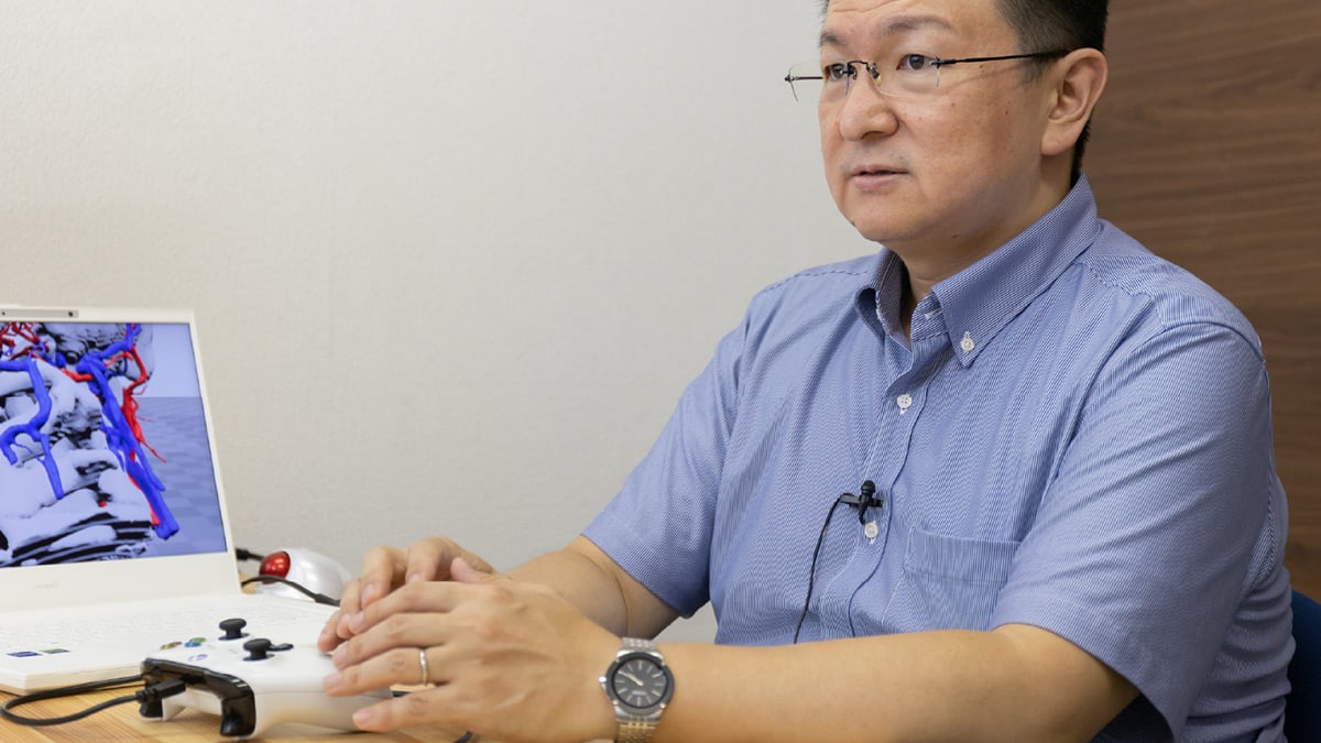
Repeated personal experiences become experiential knowledge with the development of applications
My specializations are in image processing studies, the application of VR/AR, and spatial reproduction technologies for medical and disaster prevention purposes. I have also been involved in the research of three-dimensional images with medical and dental schools for about 20 years and the application of VR/AR technologies for disaster prevention since the Great East Japan Earthquake. Furthermore, I have been developing educational support applications for students in the School of Dentistry at Kanagawa Dental University. I have been guiding graduate school students’ research on the topic of VR/AR applications in the dental field for their doctoral theses. The aim is to facilitate my personal experience for an easier understanding of difficult information to better comprehend through visualization with three-dimensional image displays. This sharing facilitates the formation of their experiments, which is why I am involved in such technical developments.
An environment that is seamlessly connected and suited for developments within the field of dentistry
I have always had an interest in the structure of the human body, so I’m incredibly grateful for the opportunity to engage in current research and education with the faculty at the School of Dentistry. The benefit of working alongside and in consultation with professionals in the field is significant and has been seamless. Working so closely with these professionals would not be possible if I was not a full-time faculty member. Kanagawa Dental University might be the only university in Japan, which has a dental school that is staffed with a full-time faculty in the field of information systems such as myself, that specializes in VR/AR. There are many institutions overseas that have full-time engineers that collaborate with medical professionals in hospitals, but these are still relatively rare in Japan. Dentistry in particular has a significant need for 3D technology, as dentists routinely handle 3D data and use CAD/CAM to fabricate coverings.
Limitations for wearing VR goggles and smart glasses
Since 2020, I have been receiving important information from faculty members at the School of Dentistry that has been important for the development of 3D educational content for dental medicine. I now realize that wearing VR head-mounted displays and smart glasses presents several hurdles in medical practice and dental schools. Ensuring these meet proper hygiene requirements, especially since the pandemic, has made attaching and detaching wearable devices time-consuming and has created some limitations.
We are embarking on the era of naked eye stereoscopic imaging!
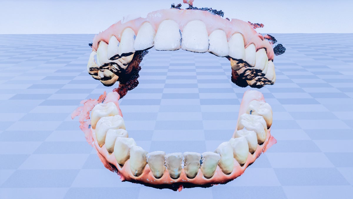
When we heard about a company that manufactured a device that could produce high-quality naked eye stereoscopic imaging in the fall of 2020, we had faculty members and students look at the application I produced. The feedback was extremely positive. By simply just sitting in front of the display, without even putting on any eyewear, images appear as if they are actual objects. Users even tried to touch displayed objects and were quite impressed. Complex structures of the human body, which were difficult to show with two-dimensional images in the past, are much easier to understand with naked eye stereoscopic imaging. It has been a highly effective educational tool. I soon realized that “we are embarking on the era of naked eye stereoscopic imaging.”
Then in 2021, Acer introduced the ConceptD 7 SpatialLabs™ Edition, a laptop equipped with technology that facilitates the experience of naked eye stereoscopic imaging. It is a personal computer that is capable of displaying naked eye stereoscopic imagery within a single unit. It’s portable, and most importantly, features an abundance of three-dimensional imaging-related applications. When this product was released in Japan in April 2022, I acquired one of the first units. I was fascinated with the product, which helped me come up with some interesting ideas. Since then, I have developed ten three-dimensional applications. Another great perk of the ConceptD 7 SpatialLabs™ Edition is that it also offers a substantial array of applications, such as “SpatialLabs™ Go”.

AI facilitates the conversion of two-dimensional video images into three-dimensional images
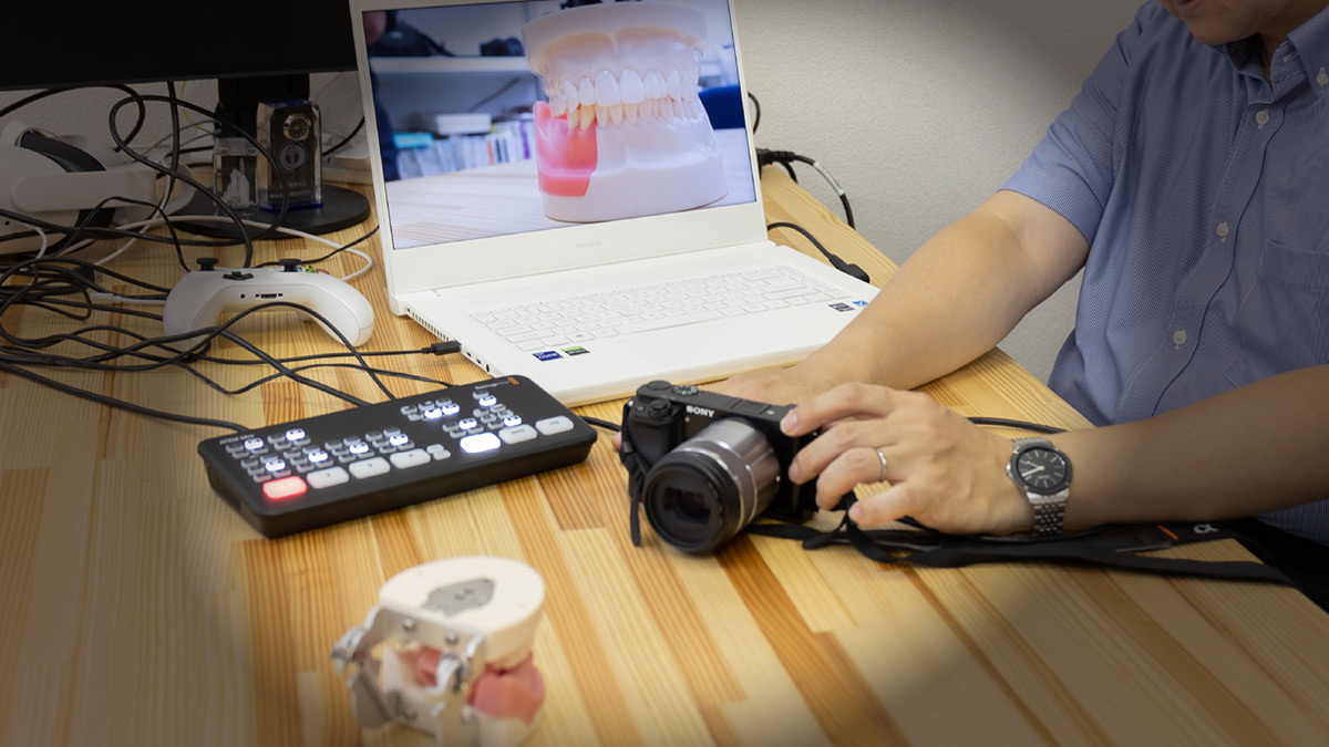
“SpatialLabs™ Go” converts live video images taken with a single lens camera into pseudo-three-dimensional images for naked eye stereoscopic imaging with AI. I believe there is a considerable demand for converting live 2D video images for stereoscopic viewing. I gave a presentation and demonstrated this application in September 2022 at the 27th Annual Conference of the Virtual Reality Society of Japan.
Live broadcasting of twin-lens camera video images requires special equipment and is quite expensive. However, I believe, that we can still learn a lot by using images captured by ordinary single-lens cameras with added depth from AI and then watching them on SpatialLabs™. There may be greater demand and broader scope for applications and methods converting videos or images taken in a training room, as opposed to stereoscopic imagery with CG models. These feature technical demonstrations performed by a faculty member using three-dimensional imagery with naked eye stereoscopic imaging on desktop displays for individual students. I have great expectations for this technology and as barriers to create content are lowered, more and more people will use it. No matter how good a device is, it will become obsolete unless it’s easy to use and content is readily available. Because of this, I see a bright future for SpatialLabs™.

The hardware is amazing, but the potential of applications is phenomenal
Dental technicians working at our university hospital asked me if it was possible to display data from ordinarily used CAD/CAM software for viewing with naked eye stereoscopic imaging. Although it isn’t currently possible to support naked eye stereoscopic imaging of data provided by unsupported software, I discovered that by distributing images through an HDMI connection that is linked to a video capturing device. By feeding that display content through the “SpatialLabs™ Go,” I could deliver pseudo-three-dimensional images for naked eye stereoscopic imaging with some degree of accuracy. This is pretty amazing, and I believe there is a need for this. So, I am trying out different methods using just standard applications. I feel there is potential for the conversion of images captured by single-lens cameras and in converting screen displays of other computers into pseudo-three-dimensional images for naked eye stereoscopic imaging using AI. Although the 2D to 3D conversion by AI is currently in development, accuracy is improving each year. It is becoming clearer which objects are easier to convert into naked eye stereoscopic imagery.
Predictions for the era for naked eye stereoscopic imaging
Acer’s new project is a game changer as it enables practical naked-eye stereoscopic viewing on a stand-alone laptop computer. We may be seeing the dawn of the naked eye stereoscopic imaging era. However, having other manufacturers enter naked eye stereoscopic imaging will help spark competition, broaden the uses, and will likely push the field further. Users will not be limited to those in medical care, and new categories for naked eye stereoscopic imaging will likely evolve. This will only further drive progress. I have high hopes for the industry, and I am convinced that it will happen in the future.
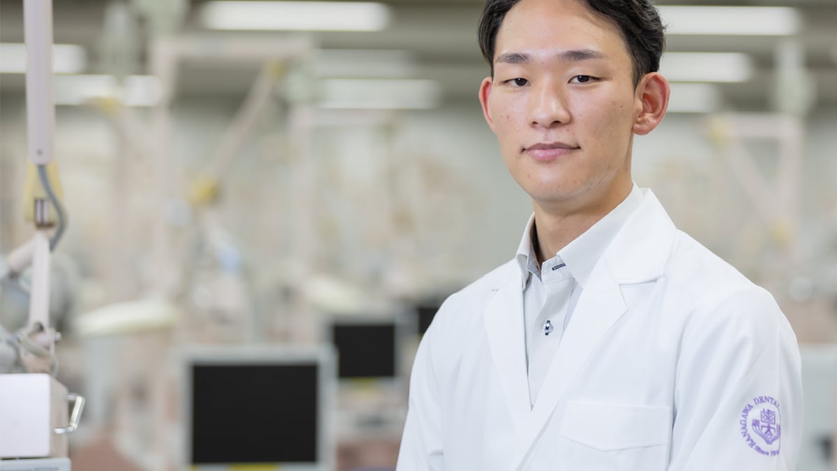
Interview with dentist and graduate student: Dr. Takahito Tsukuda
We are aiming to use digital technology to benefit research, education, and patients
How VR/AR digital technology has impacted the life of a researcher
Currently, I am working at the university hospital as a dentist while conducting and assisting in dental care. I am also doing my research at the Graduate School of Dentistry, Kanagawa Dental University. During my first year, I attended a lecture by Professor Itamiya and was struck by his introduction to VR/AR technology. This was so impactful that I changed the theme of my research, which until that time had been focused on animal experimentation. Under the guidance of Professor Itamiya, I started researching methods for applying digital technology to education. Despite the hurdles, I believe it would be wonderful if digital technology could be applied to patients in clinical settings to help provide more detailed explanations.
My reaction was, “What, it’s already available?”
I was also surprised when Dr. Itamiya introduced me to ConceptD 7 SpatialLabs™ Edition for the first time. Although I thought that someday an integrated computer with naked-eye stereoscopic viewing would be available eventually, I was amazed to see that it was already released. In a clinical setting, the examination room and operating room are in separate locations. However, having a lightweight and versatile unit in the examination room to provide explanations to patients is useful. As a bonus, it will certainly facilitate my research.

This should be useful for simulations of preoperative procedures
A microscope, cone beam computed tomography (CBCT), and computed tomography (CT) are used to explore conditions at the roots of teeth. However, these tend to rely largely on the technical acuity of practitioners. Although these may not be necessarily needed for experienced dentists, the ability to understand the conditions at the roots of the teeth stereoscopically with three-dimensional images is useful for simulating preoperative procedures for students and clinical trainees. I am also hopeful that this product proves useful as an educational tool.
Viewing stereoscopic imaging from the perspective of experienced practitioners for understanding techniques
The integration of the movements of an experienced practitioner with the movements of 3D images displayed on a screen is likely to become a useful tool for remote education in the future. However, actually showing and demonstrating the techniques of intraoral treatments to students can be challenging. There are many foreign students that come to Japan to learn about dental technology, but this may also be useful for remote classes. Learning surgical operations by observing practitioners is the most critical component of clinical study. Observing from a surgeon's point of view in three dimensions is a great learning experience for students and young doctors. Furthermore, since naked-eye three-dimensional vision can be achieved without goggles, we want to capture the techniques of skilled teachers and utilize them for education.
Interview with dental technician: Mr. Toshifumi Nakashizuka
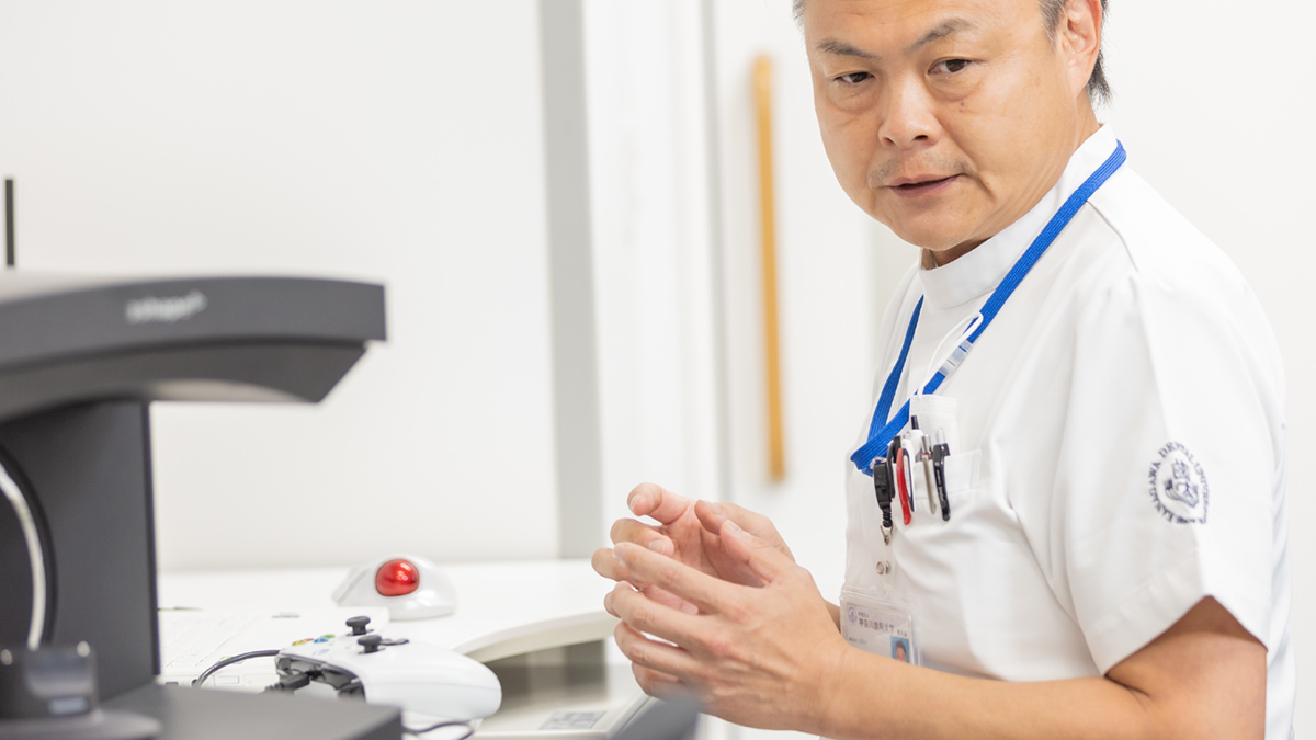
Images that seem to jump out of the screen
I’ve seen images on naked eye stereoscopic imaging displays manufactured by other companies before, but I feel 3D model images on the ConceptD 7 SpatialLabs™ Edition have smoother movements and images that seem to jump out of the screen. As a comparison, the three-dimensional images I’ve seen in the past appeared as if they were contained in a box.
I can see potential and can sense the possibilities in putting analog dental technology on a digital platform

Digitization in the field of dentistry has progressed substantially over the years, but the world of dental technicians is still analog. Technicians use their hands, which require years of experience and skills to create objects. These are made based on two-dimensional CAD data and physically fitting them on actual patients. Cosmetic treatments, particularly on front teeth, require manual adjustments to ensure that teeth look natural. They must also be adjusted to ensure they look right from a variety of angles. From this perspective, it is also nice to be able to check the image from different angles with naked-eye stereoscopic viewing. Being able to apply and fine-tune CAD/CAM data with a 3D model would be great for putting the analog dental technology we have cultivated to date on a digital platform. I see potential there.
I would like to utilize this to communicate with patients
If the dental industry decides to utilize this naked-eye stereoscopic vision in the future, it will be quite useful in providing explanations to patients. As dental technicians, we are used to conceiving 3D objects out of 2D data, but that’s not the case for our patients. Verbally explaining with 2D images does not make it any easier for them to understand what we are trying to communicate. Using naked eye stereoscopic imaging to provide explanations would make communication much more effective. Also, since it's a laptop, it's easy to transport. I have great expectations for this.
Interview with doctor of dentistry: Professor Katsuhiko Kimoto
Toward “Digital Dentistry”

Advancing dentistry with the power of digitization
Dentistry consists of dental preservation and prosthodontics. I specialize in the field of prosthodontics which is the practice of replacing missing teeth with implants, dentures, and bridges when teeth are chipped or missing. Unlike dental preservation, customizing artificial objects for individual patients is required in prosthodontics. The work dental technicians put into making crowns in collaboration with dentists takes a lot of time and effort. We are working hard to streamline this part of the process through the power of digital technology. This is the field that has been referred to recently as “digital science and technology” or “digital dentistry.”
The arrival of a new era

I was quite surprised the first time I saw the ConceptD 7 SpatialLabs™ Edition. It was interesting that you could view naked eye stereoscopic imagery on a laptop computer. Furthermore, I then realized that a new era of using 3D images in both clinical and educational settings of dentistry had arrived. Naked eye stereoscopic imaging displays were available in the past, but using these devices required replacing existing equipment. There were also other issues such as practicality. The fact that this has been achieved with a laptop computer suggests that we are getting closer to this reality.
We are aiming for a future where a unit is available for every student

It would be beneficial from an educational perspective if costs come down more in the future and each student can have their own device. The maxillofacial region, in particular, consists of fine blood vessels and nerves intertwined in a complex manner and integrated with muscles. It is extremely difficult for students to gain an understanding of this region. Currently, we’re using models to enhance understanding, but it is easier to modify a three-dimensional model than a replica. That way, it is possible to see inside the model to further enhance their understanding. I would also like to put virtual training to practical use.
A turning point for dentistry DX
Since the product is a laptop that can be easily transported, it is possible to place it near the patient when providing explanations. I would like to use this for preoperative simulations conducted by dental surgeons, to give explanations at conferences, and to raise awareness about oral hygiene. We are excited about the broad applications. I believe that the presence of ConceptD 7 SpatialLabs™ Edition will mark a turning point for dentistry DX.

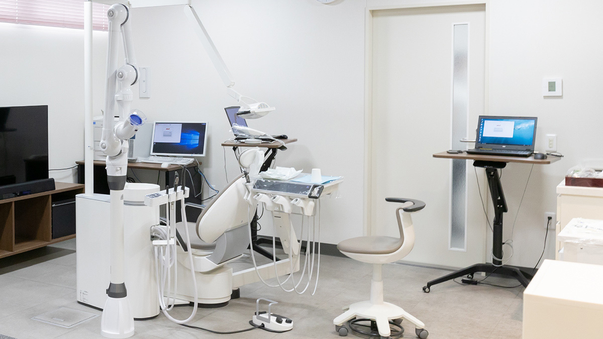
The product in use at Kanagawa Dental University/Kanagawa Dental University Hospital

ConceptD 7 SpatialLabs™ Edition “CN715-73G-SL76Z”
Disclaimer: The ConceptD 7 SpatialLabs™ Edition “CN715-73G-SL76Z” is not a medical device. The product may be used for educational and research purposes and not for examinations and treatments.